Current gold-standard histopathology for cancerous biopsies is destructive, time consuming, and limited to 2D slices, which does not faithfully represent true 3D tumor micro-morphology. Our research goal is to utilize dual-inverted Selective Plane Illumination Microscopy (diSPIM) for 3D imaging of tumors and rendering their digital histological images. By looking at the 3D volume with less sampling issues caused by physical cutting, and with the help of further computational analysis, the imaging system shows the potential for better understanding the structures of tumors, speeding up triage, and improving diagnosis. Furthermore, by adding electronic confocal slit detection (eCSD) and structured illumination (SI), diSPIM has been optimized to achieve higher image contrast (Figure A), enabling useful pseudo-H&E imaging of even highly-scattering (uncleared) tissues. Also, to improve imaging depth and cover larger 3D volume, optical clearing methods, such as TDE and CLARITY, are also being investigated. Traditional histology-like digital sections can be obtained from the reconstructed 3D volume (Figure B). By utilizing dual-view imaging and joint deconvolution, isotropic resolution over large fields of view can be achieved.
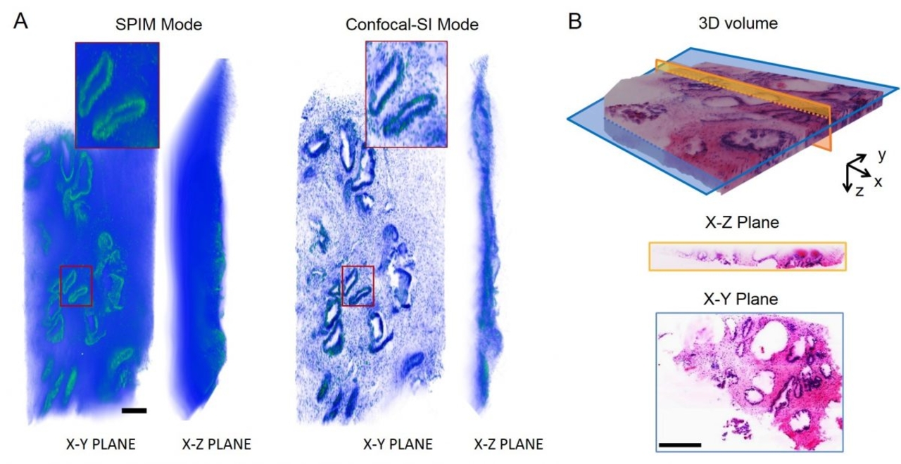
For 3D imaging of uncleared tissue, scattering background deteriorates imaging performance (Fig. A, SPIM Mode). By adding electronic confocal slit detection (eCSD, Confocal mode) and structured illumination (SI mode), imaging contrast has been tremendously improved (Fig. A, Confocal-SI Mode). As for cleared tissue with less scattering background, by imaging and reconstructing D&E stained sample at SPIM mode, traditional histology-like images can be visualized as a 3D volume or sections at arbitrary orientations (Fig. B). Scale bar: 250µm (A), 500µm (B).